One of the topics that veterinarians ask me about most frequently is how to interpret fungal cultures grown on dermatophyte test medium (DTM). The fungal culture is an important early step in working up many presentations, including folliculitis, patchy alopecia, and even facial crusting that resembles pemphigus foliaceus. As a zoonotic disease, dermatophytosis is not one you want to overlook on your differential diagnosis list.
Understanding what makes a DTM “tick” will help your interpretation. The DTM incorporates antibiotics (gentamicin and chlortetracycline) to suppress bacterial growth, cycloheximide to suppress saprophytic fungal growth, and phenol red as a color indicator. It provides both carbohydrate and protein nutrients. Dermatophytes (fungi belonging to the genera Microsporum, Trichophyton, and Epidermophyton) preferentially use protein as an energy source, whereas most saprophytic fungi use carbohydrates first. This characteristic is the key to interpreting the color change that may be seen with DTMs. The early growth of dermatophytes will usually (but not always!) cause the DTM to change from yellow to red due to the production of alkaline byproducts of protein metabolism. Saprophytes may do the same, but usually only when the colony is much larger. Check your cultures daily to make sure the red color change precedes the growth of a large colony. In addition to observing for a red color change, the interpretation of DTM growth should include an assessment of gross and microscopic colony morphology. Dermatophytes grown on DTM are generally lightly pigmented (white, buff, tan or cinnamon-colored), whereas common saprophytes are often darkly pigmented. The colony surfaces of the dermatophytes of veterinary importance are often described as powdery, granular or cottony.
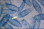
I prepare a slide for microscopic examination of suspect colonies by lightly touching a piece of clear cellophane tape to the surface of the colony, then placing it on the slide with a drop of the blue stain from a modified Wright’s kit. Macroconidia of M. canis and M. gypseum are usually easy to find, but T. mentagrophytes often doesn’t produce macroconidia on DTM. M. canismacroconidia typically have
six or more cells, thick cell walls and knobby ends. M. gypseum macroconidia tend to be numerous, ellipsoid, have thinner cell walls, and four to six cells. Some microscopic characteristics of T. mentagrophytes that can be seen are spiral hyphae and grape-like clusters of one-celled microconidia.
Consider the occurrence of a color change, the macroscopic colony appearance, and the microscopic findings into your interpretation. The fungal culture is one of the most important laboratory procedures in veterinary dermatology. It should be used to rule out ringworm, as much as to confirm it. If most of their cultures come back positive, then you may not be recommending them often enough.
Jon Plant, DVM, DACVD


